Cursiefen, Claus | Hadrian, Karina | Hos, Deniz - B 04
Targeting the osmosensitive transcription factor NFAT5 in acute and chronic corneal edema
Introduction
The cornea is the transparent avascular outer barrier and major refractive element of the eye. Loss of corneal transparency, e.g. due to dysfunction of corneal endothelial cells resulting in corneal swelling (edema), leads to corneal blindness and is the second most common cause for blindness worldwide. The only possible treatment so far is corneal transplantation, resulting in more than a million people suffering from corneal blindness due to shortage of donor corneas worldwide. Due to shortage of tissue donors, only 1 in 70 patients can be cured. Thus, non-surgical approaches to reduce corneal edema would be of great therapeutic value to treat corneal blindness. In this context, we recently discovered a novel suppressive role for the osmosensitive transcription factor nuclear factor of activated T cells 5 (NFAT5; or tonicity-responsive enhancer binding protein; TonEBP) in corneal edema resorption, thereby identifying a novel potential therapeutic target to non-surgically combat edema-induced corneal blindness. Aim of this work is to translate these findings into a clinically applicable direction by therapeutically modulating NFAT5 in the cornea in mouse models of acute and chronic corneal edema. Furthermore, this study will investigate the impact of modulating NFAT5 on graft survival after corneal transplantation.
Figure 1
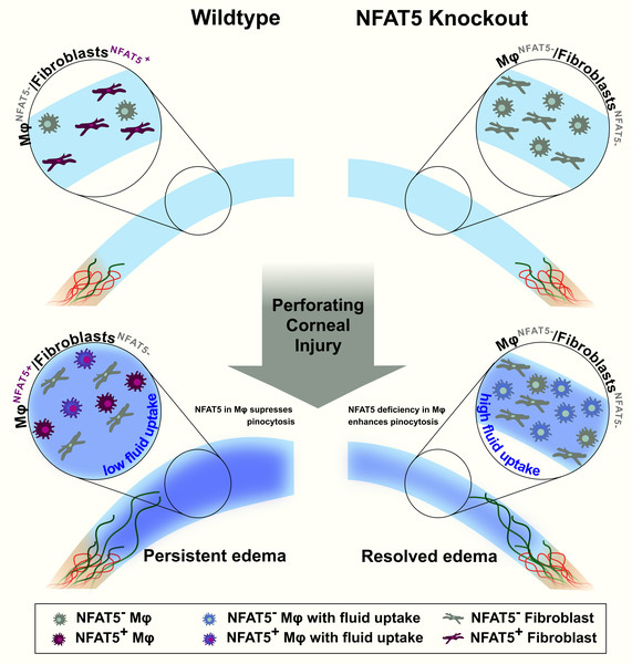
Figure 1: Presumed mechanism for the role of NFAT5 in acute corneal edema resorption after perforating corneal injury (PCI). The naive (wild-type) cornea expresses NFAT5 mainly in vimentin+ fibroblasts (purple), whereas the majority of corneal macrophages are NFAT5- (gray). Blood (red) and lymphatic (green) vessels are present at the limbus. After PCI in wild-type mice, NFAT5 is mainly expressed by corneal macrophages (purple). In wild-type corneas, macrophage pinocytosis (blue) is low, resulting in persistent corneal edema. In contrast, macrophages in mice defective for NFAT5 show higher pinocytosis capacity, leading to a faster resorption of corneal edema after PCI; lymphatic vessels are unaltered. (Hadrian et al., 2022)
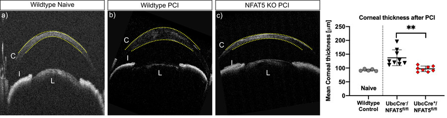
Recently, using an established murine model of PCI (“acute injury-induced corneal edema”) with a tamoxifen-inducible systemic NFAT5 knockout (UbcCre/NFAT5fl/fl) and mice with a myeloid-specific NFAT5 knockout (LysMCre/NFAT5fl/fl), we could for the first time demonstrate, that the loss of NFAT5 in uninjured corneas did not result in alterations in mean corneal thickness (MCT) as a measure of corneal edema in NFAT5 KO mice compared to wildtype mice. However, after PCI, corneas of wildtype mice showed severe edema compared to corneas of NFAT5 KO mice with an MTC comparable to uninjured corneas (Figure 2).
Figure 2
Figure 2: Corneal thickness (yellow dotted line) in naive wildtype mice (a) and one week after perforating corneal injury (PCI) in wildtype mice (b) and tamoxifen-inducible systemic NFAT5 knockout mice (c) (C: cornea; I: iris; L: lens). Modulation of NFAT5 improves corneal edema resolution.
It was previously shown that NFAT5 regulates lymphangiogenesis in the skin via macrophages. Furthermore, we have previously demonstrated that macrophage-mediated corneal lymphangiogenesis after PCI seems to influence the resorption of corneal edema (Hos et al., 2017). Interestingly, we could not detect obvious changes in the lymphatic vessel architecture in corneas of mice lacking NFAT5, indicating that lymphangiogenesis might not be the main mechanism for the increased resorption of corneal edema in this setting. Another mechanism which might be involved in the accelerated resorption of corneal edema in NFAT5 deficient mice is increased pinocytosis of macrophages. Indeed, we were able to show a significantly higher pinocytosis capacity of BMDMs generated from LysMCre+/NFAT5fl/fl mice compared to LysMCre-/NFAT5fl/fl mice. Based on these findings, we propose that loss of NFAT5 results in higher corneal macrophage numbers with increased pinocytotic capacity, leading to a faster resorption of corneal edema after injury (Hadrian et al., 2022).
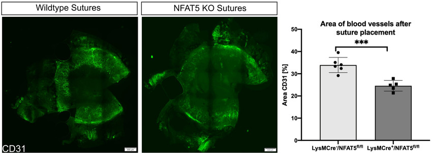
As already mentioned, we also were the first group investigating the influence of NFAT5 on corneal hemangiogenesis and tissue vascularization using an established mouse model of suture-induced inflammatory corneal neovascularization. This model leads to an ingrowth of blood as well as lymphatic vessels into the inflamed cornea. To our knowledge, the effect of NFAT5 on blood vessels also outside the eye has not been investigated yet. Our preliminary results show decreased corneal neovascularization 1 week after suture placement in mice with a myeloid-specific NFAT5 knockout (Figure 3).
Figure 3
Figure 3: Corneal vascularization visualized by CD31 staining 1 week after suture placement in wildtype mice compared to myeloid-specific NFAT5 knockout mice (LysMCre/NFAT5fl/fl) show decreased corneal neovascularization.
This is in line with a decreased expression of VEGF-A in corneas from LysMCre+/NFAT5fl/fl mice compared to LysMCre-/NFAT5fl/fl mice. The observation that NFAT5 is a regulator of corneal hemangiogenesis might be of particular importance in the setting of corneal transplantation, as it is established that inhibition of corneal hemangiogenesis promotes corneal transplant survival (Hou et al., 2018; Le et al., 2018; Hou et al., 2017).
However, for a better clinical translation, a feasible method for the local inhibition of NFAT5 at the cornea has to be established, preferably in form of eye drops. Using eye drops leads to very high local concentrations usually with few systemic side effects, has high patient compliance, and usually causes no pain. Typically, siRNAs are a common way to knock down target genes, which is also feasible in in vivo models. Therefore, we will establish an siRNA delivery system using a hybrid silicon-lipid nanoparticle system for an optimal knock down of NFAT5 at the cornea. The system contains a unique silicon-based delivery platform, which is a biocompatible hybrid of porous silicon nanoparticles and lipids (Baran-Rachwalska et al., 2020). It has been shown that this system has the ability to bind nucleic acid and deliver functional siRNA to corneal cells both in vitro and in vivo (Baran-Rachwalska et al., 2020). Therefore, we will use this system to optimize the efficacy of the siRNA knockdown of NFAT5 in the cornea.
Clinical Relevance
The main indication for corneal transplantation is the loss of corneal transparency due to severe edema. This project aims to better define the novel role of NFAT5 in corneal edema regulation and transplant immunology. Translationally, our findings may provide not only the rational but also a novel therapeutic tool to non-surgically treat corneal edema and re-establish corneal transparency and vision. Further, modulation of NFAT5 may also allow enhanced graft survival after transplantation.
Approach
Based on our own previous work and the published literature,we hypothesize that (i) local inhibition of NFAT5 by siRNA eye drops is able to reduce corneal edema, re-establish corneal transparency and reduce the need of corneal transplantation. Furthermore, we hypothesize that (ii) modulation of NFAT5 increases corneal graft survival by reducing corneal hemangiogenesis and local alloimmune responses.
In the first part, of the project, we will establish the successful in vivo siRNA knockdown of NFAT5 to reduce corneal edema. We will knock down NFAT5 in the cornea using siRNA-based eye drops. For an optimal delivery of the siRNA, hybrid silicon-lipid nanoparticles will be used.
Using this established injury model, we will now knock down NFAT5 using siRNA eye drops
In the second part of this project, the impact of the knockdown of NFAT5 on chronic corneal edema will be studied. A mouse model with a Col8a2 Q455K knock-in mutation (Col8a2Q455K/Q455K) will be used, which is already established and available in the laboratory (Jun et al., 2012; Leonard et al., 2019). This mouse line shows features strikingly similar to human FECD, including progressive alterations in endothelial cell morphology, endothelial cell loss and basement membrane guttae leading to chronic corneal edema and transparency loss. To knockdown NFAT5 in this mouse model, NFAT5 siRNA eye drops will be applied three times daily for 2 weeks. Analysis will be performed 0, 7 and 14 days after eye drops.
In the third part of the project, the impact of NFAT5 on corneal graft survival after penetrating keratoplasty (PK) will be investigated. Two established mouse models will be used for this purpose: mice with a myeloid-specific NFAT5 knockout (LysMCre/NFAT5fl/fl) and mice with a tamoxifen-inducible systemic NFAT5 knockout (UbcCre/NFAT5fl/fl). In a well-established high-risk PK model recipient mice will undergo suture placement 2 weeks prior to PK to induce vascularized high-risk recipient beds. The donor tissue will be sourced from LysMCre/NFAT5fl/fl and UbcCre/NFAT5fl/fl mice and will be transplanted into Balb/c mice. Furthermore, corneas from wildtype mice (B10.D2) will be used as donors and transplanted into LysMCre/NFAT5fl/fl and UbcCre/NFAT5fl/fl as recipients. Transplant survival will be monitored and graded weekly for 8 weeks
In the fourth part of this project, the impact of local NFAT5 knockdown using siRNA on high-risk corneal graft survival after transplantation in wildtype mice – mimicking the human high-risk setting - will be assessed. For this purpose, high-risk PK using C57Bl/6 wildtype mice as donors and Balb/c mice as recipients will be used. Three approaches using NFAT5 siRNA will be investigated to increase the graft survival after PK
3.1. Post-transplant approach: NFAT5 siRNA will be administered as eye drops in the recipient eye 3 times daily for two weeks after PK.
3.2. Pre-transplant approach: NFAT5 siRNA eye drops will be applied 3 times daily after suture placement in the recipient until PK will be performed (2 weeks)
3.3. Pre-incubation approach: the corneal graft will be pre-incubated ex vivo for 48h with NFAT5 siRNA. Afterwards, the preincubated cornea will be transplanted into high-risk recipients.
Hadrian K, Musial M, Schönberg A, Georgiev T, Küper C, Bock F, Jantsch J, Cursiefen C, Eming SA, Hos D. The Role of the Osmosensitive Transcription Factor NFAT5 in Corneal Edema Resorption after Injury. [Accepted] Exp Mol Med (2022).
Clahsen T, Hadrian K, Notara M,[...], Hos D, [...], Cursiefen C. The novel role of lymphatic vessel in the pathogenesis of ocular diseases. [Accepted] Prog Retin Eye Res (2022).
Mestanoglu M, Handel A, Cursiefen C, Hos D. Three-year follow-up of high-risk keratoplasty following fine-needle diathermy of corneal neovascularization combined with bevacizumab. Graefes Arch Clin Exp Ophthalmol (2022).
Zhang W, Schönberg A, Basset F, Hadrian K, Hos D, Becker M, Bock F, Cursiefen C. Different Murine High-Risk Corneal Transplant Settings Vary Significantly in Their (Lymph)angiogenic and Inflammatory Cell Signatures. InvestOphthalmol Vis Sci (2022).
Hadrian K, Willenborg S, Bock F, Cursiefen C, Eming SA, Hos D. Macrophage-Mediated Tissue Vascularization: Similarities and Differences Between Cornea and Skin. Front Immunol (2021).
Peckert-Maier K, Schönberg A, Wild A, Royzman D, Braun G, Stich L, Hadrian K, Tripal K, Cursiefen C, Steinkasserer A, Zinser E, Bock F. Pre-incubation of corneal donor tissue with sCD83 improves graft survival via the induction of alternatively activated macrophages and tolerogenic dendritic cells. Am J Transplant (2021).
Cursiefen C, Hos D. Cutting Edge: Novel Treatment Options Targeting Corneal Neovascularization to Improve High-Risk Corneal Graft Survival. Cornea (2021).
Le VNH, Hou Y, Bock F, Cursiefen C. Supplemental Anti Vegf A-Therapy Prevents Rebound Neovascularisation After Fine Needle Diathermy Treatment to Regress Pathological Corneal (Lymph)Angiogenesis. Sci Rep (2020).
Schaub F, Collmer M, Schrittenlocher S, Bachmann BO, Cursiefen C, Hos D. Outcome of Descemet Membrane Endothelial Keratoplasty Using Corneas from Donors >/=80 Years of Age. Am J Ophthalmol (2020).
Hos NJ, Fischer J, Hos D, Hejazi Z, Calabrese C, Ganesan R, et al. TRIM21 Is Targeted for Chaperone-Mediated Autophagy during Salmonella Typhimurium Infection. J Immunol (2020).
Hadrian K, Melkonyan H, Schlatt S, Wistuba J, Wasmuth S, Heiligenhaus A, Thanos S, Böhm MRR. Age-related distribution and potential role of SNCB in topographically different retinal areas of the common marmoset Callithrix jacchus, including the macula. Exp Eye Res (2019).
Hos D, Le VNH, Hellmich M, Siebelmann S, Roters S, Bachmann BO, et al. Risk of Corneal Graft Rejection After High-risk Keratoplasty Following Fine-needle Vessel Coagulation of Corneal Neovascularization Combined With Bevacizumab: A Pilot Study. Transplant Direct (2019).
Hos D, Matthaei M, Bock F, Maruyama K, Notara M, Clahsen T, Hou Y, Le VNH, Salabarria AC, Horstmann J, Bachmann BO, Cursiefen C. Immune reactions after modern lamellar (DALK, DSAEK, DMEK) versus conventional penetrating corneal transplantation. Prog Retin Eye Res (2019)
Kiesewetter A, Cursiefen C, Eming SA, Hos D. Phase-specific functions of macrophages determine injury-mediated corneal hem- and lymphangiogenesis. Sci Rep (2019).
König S, Hadrian K, Schlatt S, Wistuba J, Thanos S, Böhm MRR. (2019) Topographic protein profiling of the age-related proteome in the retinal pigment epithelium of Callithrix jacchus with respect to macular degeneration. J Proteomics (2019)
Büttner C, Clahsen T, Regenfuss B, Dreisow ML, Steiber Z, Bock F, Reis A, Cursiefen C. Tyrosinase Is a Novel Endogenous Regulator of Developmental and Inflammatory Lymphangiogenesis. Am J Pathol (2019).
Hou Y, Le VNH, Tóth G, Siebelmann S, Horstmann J, Gabriel T, Bock F, Cursiefen C. UV light crosslinking regresses mature corneal blood and lymphatic vessels and promotes subsequent high-risk corneal transplant survival. Am J Transplant (2018).
Ozer O, Mestanoglu M, Howaldt A, Clahsen T, Schiller P, Siebelmann S, Reinking N, Cursiefen C, Bachmann B, and Matthaei M (2022). Correlation of Clinical Fibrillar Layer Detection and Corneal Thickness in Advanced Fuchs Endothelial Corneal Dystrophy. J Clin Med11. doi:10.3390/jcm11102815.
Volatier T, Schumacher B, Cursiefen C, Notara M. UV Protection in the Cornea:Failure and Rescue. Biology (Basel). 2022 Feb 10;11(2):278. doi:10.3390/biology11020278.
Zhang W, Schonberg A, Bock F, and Cursiefen C (2022). Posttransplant VEGFR1R2 Trap Eye Drops Inhibit Corneal (Lymph)angiogenesis and Improve Corneal Allograft Survival in Eyes at High Risk of Rejection. Transl Vis Sci Technol11, 6. doi:10.1167/tvst.11.5.6.
Peil J, Bock F, Kiefer F, Schmidt R, Heindl LM, Cursiefen C, and Schlereth SL (2022). New Therapeutic Approaches for Conjunctival Melanoma-What We Know So Far and Where Therapy Is Potentially Heading: Focus on Lymphatic Vessels and Dendritic Cells. Int J Mol Sci23. doi:10.3390/ijms23031478.
Schrittenlocher S, Grass C, Dietlein T, Lappas A, Matthaei M, Cursiefen C, and Bachmann B (2022). Graft survival of Descemet membrane endothelial keratoplasty (DMEK) in corneal endothelial decompensation after glaucoma surgery. Graefes Arch Clin Exp Ophthalmol260, 1573-1582. doi:10.1007/s00417-021-05506-4.
Schrittenlocher S, Matthaei M, Bachmann B, and Cursiefen C (2022). The Cologne-Mecklenburg-Vorpommern DMEK Donor Study (COMEDOS) - design and review of the influence of donor characteristics on Descemet membrane endothelial keratoplasty (DMEK) outcome. Graefes Arch Clin Exp Ophthalmol. doi:10.1007/s00417-022-05594-w.
Volatier T, Schumacher B, Cursiefen C, and Notara M (2022). UV Protection in the Cornea: Failure and Rescue. Biology (Basel)11. doi:10.3390/biology11020278.
Zwingelberg SB, Buscher F, Schrittenlocher S, Rokohl AC, Loreck N, Wawer-Matos P, Fassin A, Schaub F, Roters S, Matthaei M, Heindl LM, Bachmann BO, and Cursiefen C (2022). Long-Term Outcome of Descemet Membrane Endothelial Keratoplasty in Eyes With Fuchs Endothelial Corneal Dystrophy Versus Pseudophakic Bullous Keratopathy. Cornea41, 304-309. doi:10.1097/ICO.0000000000002737.
Handel A, Luke JN, Siebelmann S, Franklin J, Roters S, Matthaei M, Bachmann BO, Cursiefen C, and Hos D (2022). Outcomes of deep anterior lamellar keratoplasty and penetrating keratoplasty in keratoconic eyes with and without previous hydrops. Graefes Arch Clin Exp Ophthalmol. doi:10.1007/s00417-022-05643-4.
Handel A, Siebelmann S, Luke JN, Matthaei M, Cursiefen C, and Bachmann B (2022). Influence of Body Position on Intraocular Pressure After Descemet Membrane Endothelial Keratoplasty: A Prospective Randomized Trial. Cornea. doi:10.1097/ICO.0000000000003010.
Hribek A, Mestanoglu M, Clahsen T, Reinking N, Frentzen F, Howaldt A, Siebelmann S, Bachmann BO, Cursiefen C, and Matthaei M (2022). Scheimpflug Backscatter Imaging of the Fibrillar Layer in Fuchs Endothelial Corneal Dystrophy. Am J Ophthalmol235, 63-70. doi:10.1016/j.ajo.2021.08.019.
Mestanoglu M, Handel A, Cursiefen C, and Hos D (2022). Three-year follow-up of high-risk keratoplasty following fine-needle diathermy of corneal neovascularization combined with bevacizumab. Graefes Arch Clin Exp Ophthalmol 260, 2383-2385. doi:10.1007/s00417-021-05546-w.
Roters S, Rokohl AC, Heindl LM, and Cursiefen C (2022). Novel eccentric corneoscleral donor preparation technique providing corneoscleral tectonic and central split corneal grafts for multiple recipients. Graefes Arch Clin Exp Ophthalmol 260, 2069-2071. doi:10.1007/s00417-021-05482-9.
Schaub F, Bachmann BO, and Cursiefen C (2022). Silicone oil endotamponade in eyes with Boston Keratoprosthesis Type 1. Acta Ophthalmol 100, e1041-e1042. doi:10.1111/aos.15026.
Schaub F, Mestanoglu M, Cursiefen C, and Hos D (2022). Impact of early intensified postoperative corticosteroids on immune reaction rates after Descemet membrane endothelial keratoplasty (DMEK). Graefes Arch Clin Exp Ophthalmol 260, 693-695. doi:10.1007/s00417-021-05393-9.
Hou Y, Bock F, Hos D, and Cursiefen C (2021). Lymphatic Trafficking in the Eye: Modulation of Lymphatic Trafficking to Promote Corneal Transplant Survival. Cells10. doi:10.3390/cells10071661.
Norrick A, Esterlechner J, Niebergall-Roth E, Dehio U, Sadeghi S, Schroder HM, Ballikaya S, Stemler N, Ganss C, Dieter K, Dachtler AK, Merz P, Sel S, Chodosh J, Cursiefen C, Frank NY, Auffarth GU, Ksander B, Frank MH, and Kluth MA (2021). Process development and safety evaluation of ABCB5(+) limbal stem cells as advanced-therapy medicinal product to treat limbal stem cell deficiency. Stem Cell Res Ther12, 194. doi:10.1186/s13287-021-02272-2.
Schlereth SL, Hos D, Matthaei M, Hamrah P, Schmetterer L, O'Leary O, Ullmer C, Horstmann J, Bock F, Wacker K, Schroder H, Notara M, Haagdorens M, Nuijts R, Dunker SL, Dickman MM, Fauser S, Scholl HPN, Wheeler-Schilling T, and Cursiefen C (2021). New Technologies in Clinical Trials in Corneal Diseases and Limbal Stem Cell Deficiency: Review from the European Vision Institute Special Interest Focus Group Meeting. Ophthalmic Res64, 145-167. doi:10.1159/000509954.
Handel A, Siebelmann S, Hos D, Ogrunc F, Matthaei M, Cursiefen C, and Bachmann B (2021). Comparison of Mini-DMEK versus predescemetal sutures as treatment of acute hydrops in keratoconus. Acta Ophthalmol99, e1326-e1333. doi:10.1111/aos.14835.
Hayashi T, Zhang W, Hos D, Schrittenlocher S, Nhat Hung Le V, Siebelmann S, Matthaei M, Bock F, Bachmann B, and Cursiefen C (2021). Descemet Membrane Endothelial Keratoplasty in Vascularized Eyes: Outcome and Effect on Corneal Neovascularization. Cornea40, 685-689. doi:10.1097/ICO.0000000000002502.
Hribek A, Mestanoglu M, Clahsen T, Reinking N, Frentzen F, Howaldt A, Siebelmann S, Bachmann BO, Cursiefen C, and Matthaei M (2021). Scheimpflug Backscatter Imaging of the Fibrillar Layer in Fuchs Endothelial Corneal Dystrophy. Am J Ophthalmol235, 63-70. doi:10.1016/j.ajo.2021.08.019.
Schrittenlocher S, Grass C, Dietlein T, Lappas A, Matthaei M, Cursiefen C, and Bachmann B (2021). Graft survival of Descemet membrane endothelial keratoplasty (DMEK) in corneal endothelial decompensation after glaucoma surgery. Graefes Arch Clin Exp Ophthalmol. doi:10.1007/s00417-021-05506-4.
Zhang W, Schonberg A, Hamdorf M, Georgiev T, Cursiefen C, and Bock F (2021). Preincubation of donor tissue with a VEGF cytokine trap promotes subsequent high-risk corneal transplant survival. Br J Ophthalmol. doi:10.1136/bjophthalmol-2021-319745.
Cursiefen C, and Hos D (2021). Cutting Edge: Novel Treatment Options Targeting Corneal Neovascularization to Improve High-Risk Corneal Graft Survival. Cornea40, 1512-1518. doi:10.1097/ICO.0000000000002736.
Hadrian K, Willenborg S, Bock F, Cursiefen C, Eming SA, and Hos D (2021). Macrophage-Mediated Tissue Vascularization: Similarities and Differences Between Cornea and Skin. Front Immunol12, 667830. doi:10.3389/fimmu.2021.667830.
Hos, D., Matthaei, M., Bock, F., Maruyama, K., Notara, M., Clahsen, T., Hou, Y., Le, V.N.H., Salabarria, A.C., Horstmann, J., Bachmann, B.O., and Cursiefen, C. (2019). Immune reactions after modern lamellar (DALK, DSAEK, DMEK) versus conventional penetrating corneal transplantation. Prog Retin Eye Res10.1016/j.preteyeres.2019.07.001.
Notara M, Lentzsch A, Clahsen T, Behboudifard S, Braun G, Cursiefen C: Bevacizumab Induces Upregulation of Keratin 3 and VEGFA in Human Limbal Epithelial Cells in Vitro. J Clin Med. 2019 Nov 9;8(11):1925. doi: 10.3390/jcm8111925. PMID: 31717500
Clahsen T, Buttner C, Hatami N, Reis A, and Cursiefen C (2020). Role of Endogenous Regulators of Hem- And Lymphangiogenesis in Corneal Transplantation. J Clin Med 9.
Hayashi T, Schrittenlocher S, Siebelmann S, Le VNH, Matthaei M, Franklin J, Bachmann B, and Cursiefen C (2020a). Risk factors for endothelial cell loss after Descemet membrane endothelial keratoplasty (DMEK). Sci Rep-Uk 10.
Le VNH, Hos D, Hou Y, Witt M, Barkovskiy M, Bock F, and Cursiefen C (2020a). VEGF TrapR1R2 Suspended in the Semifluorinated Alkane F6H8 Inhibits Inflammatory Corneal Hem- and Lymphangiogenesis. Translational vision science & technology 9, 15.
Le VNH, Hou Y, Bock F, and Cursiefen C (2020b). Supplemental Anti Vegf A-Therapy Prevents Rebound Neovascularisation After Fine Needle Diathermy Treatment to Regress Pathological Corneal (LYMPH)Angiogenesis. Sci Rep 10, 3908.
Salabarria AC, Koch M, Schonberg A, Zinser E, Hos D, Hamdorf M, Imhof T, Braun G, Cursiefen C, and Bock F (2020). Topical VEGF-C/D Inhibition Prevents Lymphatic Vessel Ingrowth into Cornea but Does Not Improve Corneal Graft Survival. J Clin Med 9.
Schaub F, Collmer M, Schrittenlocher S, Bachmann BO, Cursiefen C, and Hos D (2020). Outcome of Descemet Membrane Endothelial Keratoplasty Using Corneas from Donors >/=80 Years of Age. Am J Ophthalmol 211, 200-6.
Schrittenlocher S, Schlereth SL, Siebelmann S, Hayashi T, Matthaei M, Bachmann B, and Cursiefen C (2020). Long-term outcome of descemet membrane endothelial keratoplasty (DMEK) following failed penetrating keratoplasty (PK). Acta Ophthalmol 10.1111/aos.14417.
Siggel R, Schroedl F, Dietlein T, Koch KR, Platzl C, Kaser-Eichberger A, Cursiefen C, and Heindl LM (2020). Absence of lymphatic vessels in non-functioning bleb capsules of glaucoma drainage devices. Histology and histopathology 10.14670/HH-18-300, 18300.
This website uses cookies. Those have two functions: On the one hand they are providing basic functionality for this website. On the other hand they allow us to improve our content for you by saving and analyzing anonymized user data. You can redraw your consent to using these cookies at any time. Find more information regarding cookies on our Data Protection Declaration and regarding us on the Imprint.