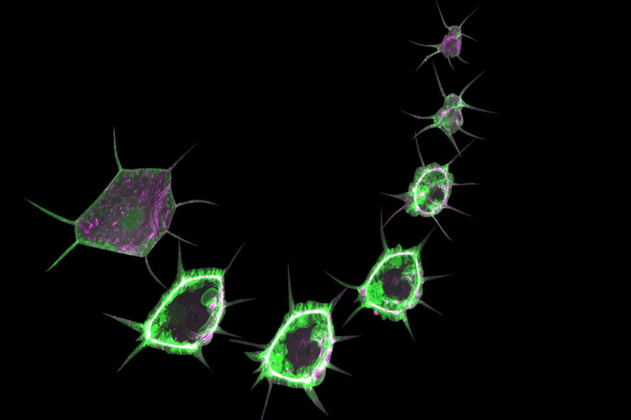
This image is a composition of snapshots from a timelapse video of a closing wound in the epidermis of a Drosophila larva. The cells express two fluorescently labelled markers: a PIP3 sensor (green) and the myosin light chain (magenta). Credit: Parisa Kakanj/EMBL
Scientists from the European Molecular Biology Laboratory (EMBL), the Institute of Genetics, CECAD and Center for Molecular Medicine Cologne (CMMC) at the University of Cologne and the Max Planck Institute for the Biology of Aging have developed a method to observe cells in the living fruit fly larvae and follow cellular processes. This technique, which has been published in the journal "Nature Protocols", offers a simple but effective way to study the functions of organs in living animals.
The fruit fly Drosophila is considered an ideal model organism in genetic research. Key processes and 60 percent of the genes of the fruit fly are identical to those of humans. Therefore, scientists have created many genetic techniques to study molecular processes of development and human diseases in the fly.
Most studies have been carried out on the embryo or the adult fly, but the fruit fly larvae also offer enormous research potential. Its transparent body with functioning organs - such as brain, intestines and muscles - makes the larva an excellent object of research for observing the dynamics of cells and molecular processes in living animals, explains Dr. Parisa Kakanj from the Cologne Institute of Genetics, who headed the study: "The cells in a fruit fly larva are much larger than those in the embryo. Therefore, all organelles and even the smallest processes can be seen. That's the beauty of this system."
Some molecular processes and interactions in a cell can only be deciphered by long-term observation. However, the continuous crawling of the larva makes such observation difficult. Kakanj has taken up this challenge and developed a method for easy immobilization of the larvae. This enables long-term live observations of cells in high resolution.
In the past, scientists used mechanical methods or anaesthetics to immobilize fruit fly larvae. However, both approaches had undesired side effects. In addition, simultaneous microscopic imaging of many larvae was not possible. "In contrast to previous techniques, we have developed a simple method for short-term treatment with ether, a classical anaesthetic. This approach enabled problem-free long-term immobilization of many larvae," explains Parisa Kakanj.
In order to prove the efficiency of the technique, the team investigated the wound healing of fruit fly larvae. They analyzed the influence of insulin and TOR - a crucial signal molecule for survival, growth and reproduction - on this process. In their study, the scientists created a map showing the temporal sequence of insulin and TOR signalling pathway activity during wound healing. They found that a reduction in insulin signalling activity at the wound margin slows down the healing processes.
Long-term live microscopic imaging coupled with genetic manipulation opens the way to new aspects of biology and physiology: "This technique will help many scientists to investigate neuronal signal transmission, fat metabolism or tumour formation and open up new possibilities for drug development," said Kakanj.
Scientific contact:
Dr. Parisa Kakanj
Institute for Genetics University of Cologne
+49-221 470-3528
pkakanj[at]uni-koeln.de
Press and communications:
Frieda Berg
+49 221 470-1704
f.berg[at]uni-koeln.de
Original publication:
Kakanj, P., Eming, S.A., Partridge, L. et al. Long-term in vivo imaging of Drosophila larvae. Nat Protoc (2020).
Copyright ©
Prof. Dr. med. Thomas Benzing
Chair of the CMMC