Retinal microglia and their immunological effects in AAV-based ocular gene therapy
Introduction
Gene therapies using adeno-associated viruses (AAVs) are among the most promising strategies to treat inherited retinal diseases. Although AAV vectors are considered to be safe, they are recognized as foreign invaders and elicit host-cell responses even in immune privileged areas such as the eye. A deep understanding of these immune processes in the retina is important for eliminating immune-related side effects and toxicity that negatively impact gene therapy. Microglia, the resident immune cells of the retina, are not only bystanders but can also trigger retinal degeneration and potentially serve as targets for therapy. Our preliminary data together with recent findings from collaborators indicate that microglia are early responders to locally injected AAVs. Beside the impact of virus titer on the immunogenicity of AAVs, capsid protein variants, driving promoters and the transgene product also seem to be decisive factors. Our hypothesis is that microglia acquire a cytotoxic phenotype after AAV transduction that results in chronic inflammation and reduced transgene expression. In this project, we will perform an in-depth analysis of retinal microglia at different stages after AAV transduction in response to different titers, capsid variants, ubiquitous and endogenous promoters as well as vectors containing different transgenes. We also aim to perform an experimental therapy in retinoschisin (Rs1h)-deficient mice under conditions of microglia modulation using minocycline. This study will not only help to decipher toxic mechanism and pinpoint triggers for harmful retinal immune responses but also help to elucidate whether immunomodulation is a beneficial approach for improving gene therapy efficacy and durability.
Figure 1
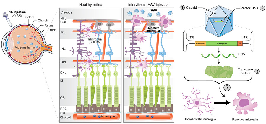
Figure 1: Intravitreal injections of recombinant adeno-associated viruses (rAAVs) trigger microglia activation, resulting in migration and accumulation of these cells in the inner retina, particularly in the ganglion cell layer (GCL). Immunogenic components of rAAV that could influence the microglial immune response are (1) AAV capsid variants, (2) vector DNA including the promoter and/or the transgene sequence and (3) the transgene protein. Identifying the factors that determine the immunogenicity of the rAAVs is crucial to achieve long-term expression of the therapeutic gene. BM, Bruch’s membrane; OS, outer segment; IS, inner segment; ONL, outer nuclear layer; OPL, outer plexiform layer; INL, inner nuclear layer; IPL, inner plexiform layer and NFL, nerve fiber layer.
Clinical Relevance
Severe ocular inflammation occurring in viral gene therapy of the retina, triggered by innate and adaptive immune responses to AAVs and cargo transgenes may build substantial hurdles to the clinical development in progress for many visually impaired patients. The identification of immune sensing triggers and potential molecular targets for eliminating immune-related side effects that impact gene therapy will help to understand the nature of inflammation and to improve interventions during gene therapy to control or prevent inflammation in patients with inherited retinal degenerations.
Approach
Aim 1: Analyze and understand retinal microglia responses after AAV2 transduction related to vector titers, capsid variants, promoters and transgenes.
Figure 2: C57BL/6J mice will be intravitreally (ivt.) injected with rAAV2 that differ in their capsid (7m8 vs GL), promoter (CAG vs Rs1) and transgene (EGFP vs. Rs1) at three different vector titers. Early and late phase microglia responses will be analyzed.
Methods:
Optomotor response to assess visual acuity
OCT and FFA to determine retinal thickness, structure and vascular integrity
IHC of retinal cryosections and flat mounts to assess:
structural changes of the retina (anti-Cone arrestin, Rho and ZO-1) and cell death (TUNEL)
microglia reactivity and morphology (anti-Iba1)
transduction efficacy of Rs1 and EGFP (+ WB)
qPCR and ELISA to assess inflammatory marker expression
RNA-Seq of isolated retinal microglia and pathway analysis
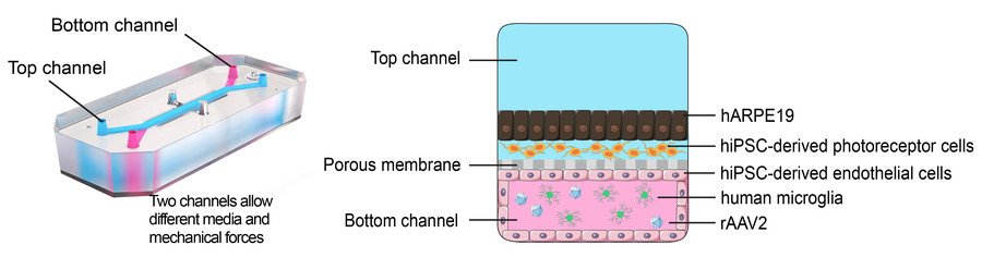
Aim 2: Analysis of microglia reactivity and validation of AAV retinal gene therapy vectors on a human-based retina-on-a-chip as a new translational model.
Figure 3: Human-based retina-on-a-chip. Schematics showing the hiPSC-derived photoreceptor cells and hARPE19 cells in the top channel and the hiPSC-derived endothelial cells and human microglia in the bottom channel. The two-channel structure allows the cells to subject to different media and fluid shear stress caused by media flow. The two channels are separated by a porous membrane.
Methods:
Live-cell imaging and immunostainings to assess:
rAAV transduction efficacy
Microglia reactivity and morphology
Blood retinal barrier penetration and cell migration
qPCR and ELISA to analyze inflammation and pathways based on findings from Aim1
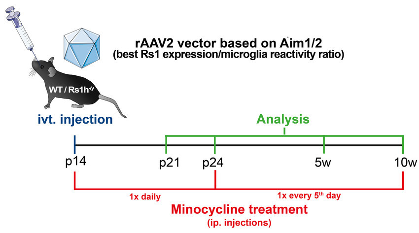
Aim 3: To perform an experimental AAV2 vector-based gene augmentation therapy in Rs1h-deficient mice under conditions of potent immunomodulation using minocycline.
Rs1h-/y mice and WT littermates will be intravitreally (ivt.) injected with rAAV2 identified in Aim1/2. Additionally, mice receive intraperitoneal injections of minocycline constantly during the gene therapy phase.
Pavlou, M. et al. Novel AAV capsids for intravitreal gene therapy of photoreceptor disorders. EMBO Mol Med 13, e13392, doi:10.15252/emmm.202013392 (2021).
Wolf, A., Herb, M., Schramm, M. & Langmann, T. The TSPO-NOX1 axis controls phagocyte-triggered pathological angiogenesis in the eye. Nat Commun 11, 2709, doi:10.1038/s41467-020-16400-8 (2020).*
Xiong, W. et al. AAV cis-regulatory sequences are correlated with ocular toxicity. Proc Natl Acad Sci U S A 116, 5785-5794, doi:10.1073/pnas.1821000116 (2019).
Xue, K. M. et al. Beneficial effects on vision in patients undergoing retinal gene therapy for choroideremia. Nat Med 24, 1507-+, doi:10.1038/s41591-018-0185-5 (2018).
Ramachandran, P. S. et al. Evaluation of Dose and Safety of AAV7m8 and AAV8BP2 in the Non-Human Primate Retina. Hum Gene Ther 28, 154-167, doi:10.1089/hum.2016.111 (2017).
Reichel, F. F. et al. AAV8 Can Induce Innate and Adaptive Immune Response in the Primate Eye. Mol Ther 25, 2648-2660, doi:10.1016/j.ymthe.2017.08.018 (2017).
Weber, B. H. et al. Inactivation of the murine X-linked juvenile retinoschisis gene, Rs1h, suggests a role of retinoschisin in retinal cell layer organization and synaptic structure. Proc Natl Acad Sci U S A 99, 6222-6227, doi:10.1073/pnas.092528599 (2002).
Chan, Y. K. et al. Reducing AAV-Mediated Immune Responses and Pathology in a Subretinal Pig Model by Engineering the Vector Genome. Molecular Therapy 27, 298-298 (2019).
Dannhausen, K., Mohle, C. & Langmann, T. Immunomodulation with minocycline rescues retinal degeneration in juvenile neuronal ceroid lipofuscinosis mice highly susceptible to light damage. Dis Model Mech 11, doi:10.1242/dmm.033597 (2018).*
Khabou, H., Cordeau, C., Pacot, L., Fisson, S. & Dalkara, D. Dosage Thresholds and Influence of Transgene Cassette in Adeno-Associated Virus-Related Toxicity. Hum Gene Ther 29, 1235-1241, doi:10.1089/hum.2018.144 (2018).
Gootwine, E. et al. Safety and Efficacy Evaluation of rAAV2tYF-PR1.7-hCNGA3 Vector Delivered by Subretinal Injection in CNGA3 Mutant Achromatopsia Sheep. Hum Gene Ther Clin Dev 28, 96-107, doi:10.1089/humc.2017.028 (2017).
Ghazi, N. G. et al. Treatment of retinitis pigmentosa due to MERTK mutations by ocular subretinal injection of adeno-associated virus gene vector: results of a phase I trial. Hum Genet 135, 327-343, doi:10.1007/s00439-016-1637-y (2016).
Karlstetter, M. et al. Retinal microglia: just bystander or target for therapy? Prog Retin Eye Res 45, 30-57, doi:10.1016/j.preteyeres.2014.11.004 (2015).*
Dalkara, D. et al. In vivo-directed evolution of a new adeno-associated virus for therapeutic outer retinal gene delivery from the vitreous. Sci Transl Med 5, 189ra176, doi:10.1126/scitranslmed.3005708 (2013).
Gehrig, A. et al. Genome-wide expression profiling of the retinoschisin-deficient retina in early postnatal mouse development. Invest Ophthalmol Vis Sci 48, 891-900, doi:10.1167/iovs.06-0641 (2007).*
Langmann, T. Microglia activation in retinal degeneration. J Leukoc Biol 81, 1345-1351, doi:10.1189/jlb.0207114 (2007).* *own publications
Prof. Dr. Thomas Langmann
Clinic of General Ophthalmology | Lab. for Experimental Immunology of the Eye
Office Anja Volkmann Lab head Dr. Anne Wolf PostDocs Dr. Sarva Keihani Dr. Mona Tabel PhD students Urbanus Kinuthia Mandy Hector Nils Laudenberg Julia Hofmann Justus Dick Master student Anna Gröger Medical students Leo Bahlmann Melis Cansu Cömert Technician Eva Scheiffert
This website uses cookies. Those have two functions: On the one hand they are providing basic functionality for this website. On the other hand they allow us to improve our content for you by saving and analyzing anonymized user data. You can redraw your consent to using these cookies at any time. Find more information regarding cookies on our Data Protection Declaration and regarding us on the Imprint.